Study in mice shows how chikungunya virus may cause joint pain
PTI, Aug 30, 2019, 1:55 PM IST

Washington: A new method for permanently marking cells infected with chikungunya virus could reveal how joint pain from the disease persists for months to years after the initial infection, says a study published in the science journal PLOS Pathogens.
Chikungunya is an arbovirus – a term used to describe viruses spread by insects – and its genetic component is the single-stranded RNA (ssRNA), as opposed to the double-stranded DNA.
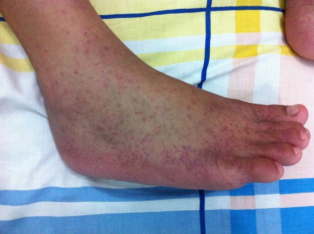
The virus is spread by Aedes aegypti and Aedes albopictus mosquitoes, and the disease usually starts with sudden fever, chills, headache, nausea, vomiting, joint and muscular pain, and rashes.
“There is neither any vaccine nor drugs available to cure the chikungunya, and the cases are managed symptomatically”, mentions the 2017-18 Annual Report by the Ministry of Health and Family Welfare, India.
According to the study’s authors – Deborah Lenschow of Washington University School of Medicine in St. Louis, USA, and colleagues – uncovering the mechanisms for chronic pain due to the disease could help in the development of treatments and preventative measures for this debilitating, virally induced arthritis.
First recognised in Tanzania in the 1950s, the term ‘Chikungunya’ originated from an ethnic phrase that means ‘to walk bend-over’ – perfectly capturing the incapacitating joint pain suffered by infected individuals.
The disease has now been reported in more than 40 countries, and approximately 30 to 60 per cent of the people infected with the virus continue to experience joint pain for months to years after the initial infection, the study says.
However, it is still not known how the joint pain persists after the infection is cured, as the replicating virus cannot be detected during the chronic phase. Seeking to answer this question, Lenschow and her colleagues developed a system to permanently mark and report on the cells infected by the arbovirus.
Using this reporter system, Lenschow and her team showed in mice that marked cells surviving chikungunya virus infection are a mixture of muscle and skin cells, present for at least 112 days after the initial virus inoculation.
When the research team treated the mice with an antibody that blocks the viral infection, it reduced the number of marked cells in the muscle and skin. The surviving marked cells also contained most of the persistent chikungunya virus RNA.
These findings provide further evidence that cells in the musculoskeletal system are targets of the chikungunya virus during the acute and chronic stages of disease.
And according to the authors, this marker-reporter system can be a useful tool for identifying and isolating cells that harbor chronic viral RNA in order to study the mechanisms behind the disease.
“Persistent CHIKV (chikungunya virus) RNA can be detected in human and animal models but no one has been able to identify where the RNA resides due to insensitive techniques,” Lenschow said. “Using our reporter system we have demonstrated that cells can survive CHIKV infection, and these cells harbor most of the persistent RNA, she said.
Commenting on the research, Krishnan H Harshan, who studies the Hepatitis C Virus at the CSIR- Centre for Cellular and Molecular Biology (CCMB), Hyderabad told PTI that the mechanism of persistence of the viral RNA in mice may not be completely reflective of the same in humans.
Harshan said that some viruses that infect humans do not have the same effect in other organisms, and that one may have to carry out similar studies in model organisms that are closer to the human physiology.
“Carrying out studies involving recombinant genetic material in such organisms is much more difficult with several ethical barriers as well,” he added.
According to Harshan, the methodology used in the current study can be seen as a prototype, extendable to understand the mechanism of persistent or resurgent infections in other viruses. However, he said that the method still does not let us see the chronological order in which different tissues in an animal’s body are infected by replicating viruses.
He explained that in viral infections, there are primary cells that the virus infects, and a lot of times, the virus replicates and then spreads into other parts of the body from these.
“Some viruses, HIV for example, persists in some later infected cells and tissues, and comes out to infect the host when we think the person is recovering. The method used in this study can be extended to understand in which cells or tissues the remaining infectious viral particles remain before emerging again to cause disease, or remain to cause persisting problems,” Harshan said.
Since many believe that the persistent RNA contributes to chronic arthritis, Lenschow added, the reporter system could be a useful tool to study the mechanisms underlying chronic disease.
Udayavani is now on Telegram. Click here to join our channel and stay updated with the latest news.
Top News

Related Articles More

High nitrate levels in groundwater threaten public health in 440 districts: Report

Gujarat IMA opposes ‘mixopathy’ proposal; says it poses ‘severe risks’ to people’s health

Study links social inequality to dementia-related changes in brain
People single all their lives might have low life satisfaction: Study

Drinking tea, coffee linked to lower risk of head and neck cancer: Study
MUST WATCH
Latest Additions

Bus Fare Hike: Government can’t keep giving subsidies endlessly, says Minister Cheluvarayaswamy

Robbers loot Rs 12 lakh jewellery from Delhi showroom, flee after being attacked with chilli powder

Started setting new goals for 2025 after World Championship and Khel Ratna: Gukesh

Delhi Police nabs man wanted in robbery case after 14-year hunt

Karkala: Couple rescued after falling into open well
Thanks for visiting Udayavani
You seem to have an Ad Blocker on.
To continue reading, please turn it off or whitelist Udayavani.


